
Brains of Congenitally Deaf Reveal Plasticity of Auditory Cortex

The human brain, when deprived of certain input for a period of time, shows a great deal of plasticity, reorganizing itself to more effectively process the input that it does receive. Research has shown, for example, that people who are born blind are often more sensitive to differences in auditory pitch and touch than people who are sighted. Similarly, studies have shown that individuals who are born deaf may be better at detecting motion and seeing in their periphery than those who can hear.
A neuroimaging study led by psychological scientist Jorge Almeida of the University of Coimbra, Portugal shows that, in people who are born deaf, the auditory cortex processes information about the visual properties of stimuli.
The research was conducted by an international team of researchers from the University of Coimbra, Beijing Normal University, Peking University, the University of Minho, and the University of Rochester. The researchers presented 10 congenitally deaf and 10 hearing Chinese participants with different visual stimuli while their brain activity was measured via functional MRI. Based on participants’ brain responses to the visual stimuli – rotating wedges and expanding annuli, shown in the figure – the researchers were able to create field maps, identifying where in the brain specific visual features were processed.
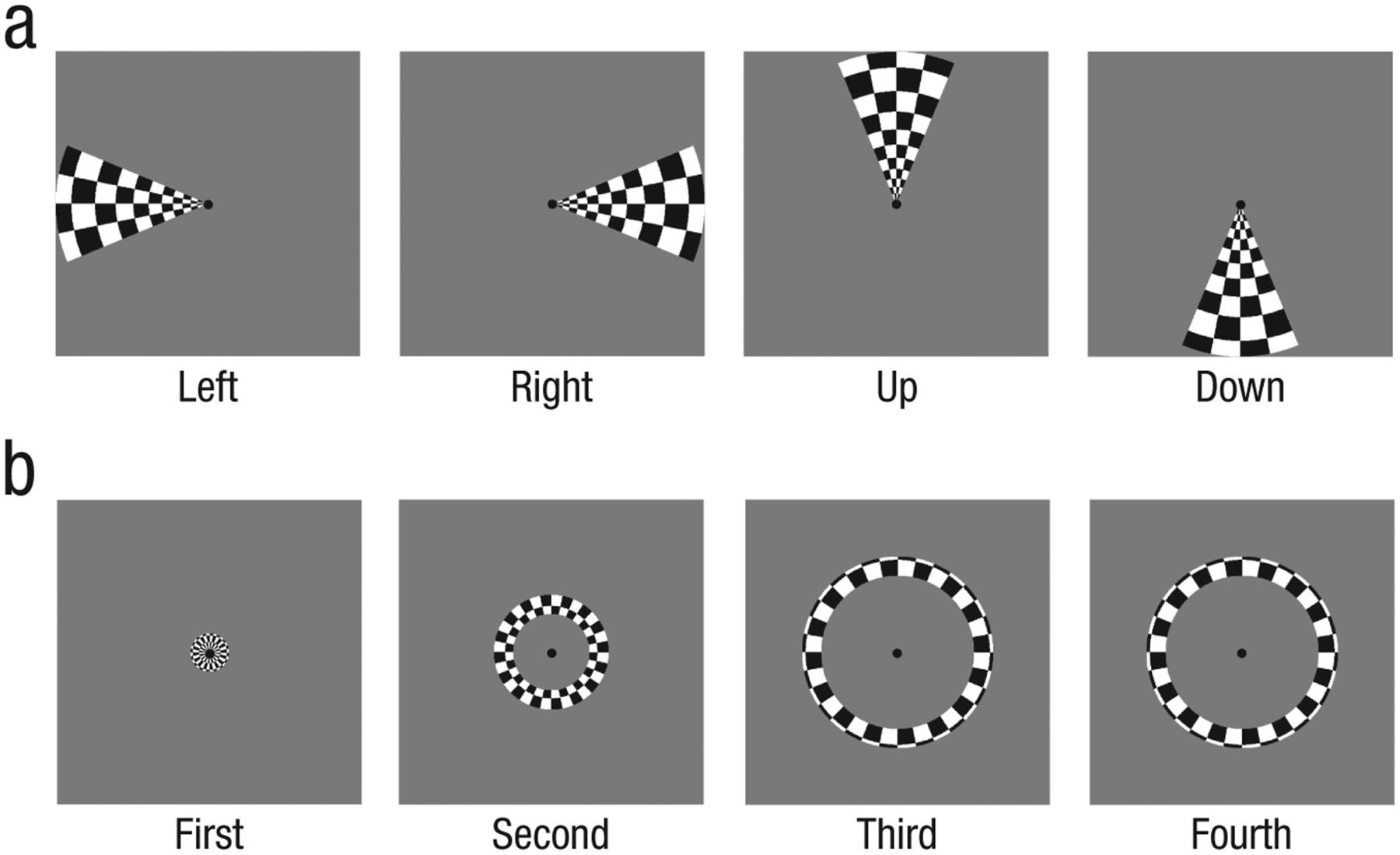
Wedges (a) and annuli (b) stimuli used in the experiment.
The results showed that, in the case of congenitally deaf individuals, the auditory cortex seemed to have taken over some of the processes typically seen within the visual cortex.
For example, the researchers were able to use activity in the auditory cortex to classify the horizontal location of a visual stimulus. And they could also use the information to classify whether a stimulus was presented in the center of the screen or in the visual periphery.
“These changes may support the enhanced visual processing in the periphery that is observed for deaf individuals,” says Almeida. “Understanding how the nervous system can ‘reprogram’ itself, and change its functional and processing properties, is essential for the development of models of long-term neuroplasticity that will eventually lead to the development of evidence-based therapies.”
According to Almeida, these findings may be especially relevant in developing therapeutic approaches for brain rehabilitation to treat trauma and neurodegenerative diseases. And it may also aid in the development of improved cochlear implants.
“Current devices explore the typical organization of the auditory system, but this study demonstrates that the auditory system is reorganized in congenital deafness, in that it represents visual information such as the location of objects in the visual field,” Almeida explains. “Thus, it may be necessary to look at how cochlear implants can exploit functional organization differently.”
Reference
Almeida, J., He, D., Chen, Q., Mahon, B.Z., Zhang, F., Gonçalves, Ó.F., Fang, F., & Bi, Y. (2015). Decoding visual location from neural patterns in the auditory cortex of the congenitally deaf. Psychological Science. DOI: 10.1177/0956797615598970

APS regularly opens certain online articles for discussion on our website. Effective February 2021, you must be a logged-in APS member to post comments. By posting a comment, you agree to our Community Guidelines and the display of your profile information, including your name and affiliation. Any opinions, findings, conclusions, or recommendations present in article comments are those of the writers and do not necessarily reflect the views of APS or the article’s author. For more information, please see our Community Guidelines.
Please login with your APS account to comment.